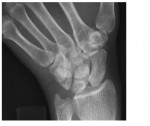
These wrist x-rays are from a 43 year old with ulnar sided wrist pain following a fall on the outstretched left hand. What can you observe?
[peekaboo_link name=”Answer”]Answer[/peekaboo_link] [peekaboo_content name=”Answer”]The oblique wrist x-ray shows a minimally displaced pisiform fracture. The x-rays taken are, in fact, scaphoid views. A pisiform fracture can be missed on standard frontal and lateral wrist views. The pisiform bone is best seen in the semi supinated oblique view.
Ulnar neuropathy is a rare complication as pisiform forms the medial border of the tunnel of Guyon which contains the ulnar artery and nerve.
If associated hamate/triquetral fractures are suspected, CT should be requested for further characterisation.
Treatment is conservative and involves a below elbow cast immobiliation and referral to a hand surgeon/orthopaedician.
References: www.wheelessonline.com
[/peekaboo_content]


